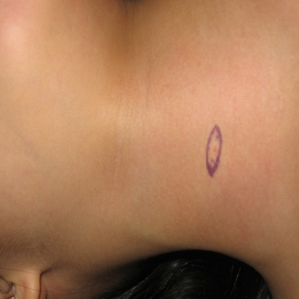
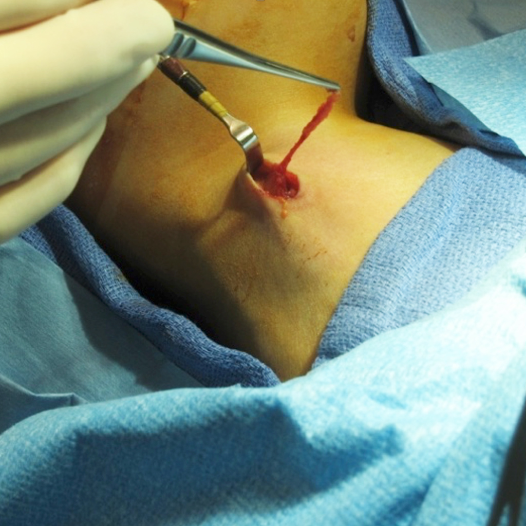
Branchial Cleft Anomalies are the result of incomplete closure of clefts or pouches in our head and neck region during embryological development. This can form in a combination of sinus tracts, fistula tracts, or cysts. A sinus is where there is one opening either on the skin or inside in the mucosa in the aerodigestive tract. A fistula is where there are two openings one on the outside in the skin and one on the inside in the mucosa. A cyst is a retained space that is part of a sinus or fistula that results in a fluid filled mass in the head and neck region. There are 4 different types of branchial cleft anomalies all based on where they originate from in the neck.

First Branchial Cleft Anomalies
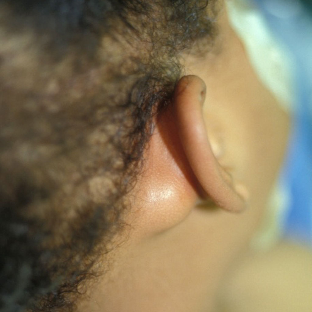
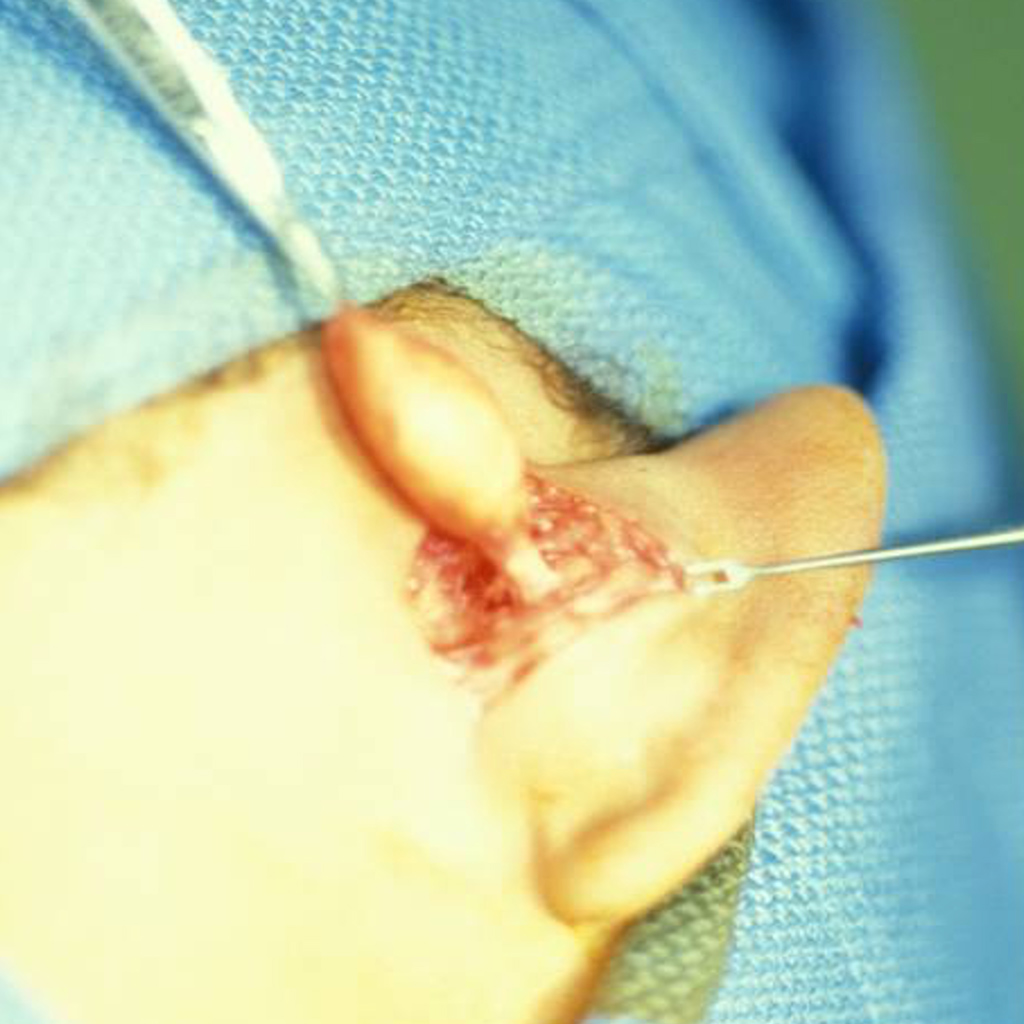
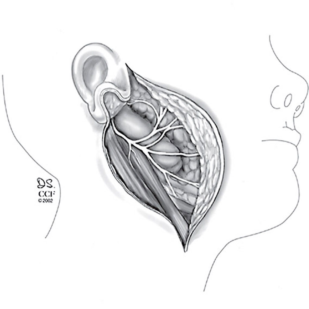
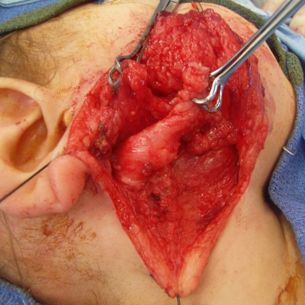
There are two types of first branchial cleft anomalies, type 1 and type 2. Both types represent a duplication of our ear canal (external auditory canal). The type 1 anomalies produce a tract parallel to our ear canal and end in the area behind our ear (Figure 1 and 2). They can often present as a cyst in this area. Type 2 anomalies are also a duplication of the ear canal but are perpendicular to the ear canal and can end in a pit or a cyst near the jaw bone (Figure 1 and 3). Both types of tracts or cysts are closely related to the facial nerve.

Second Branchial Cleft Anomalies
Second Branchial Cleft Anomalies are the most common branchial cleft anomalies. They can present as a mass in the antero-lateral part of the neck either early or later in life, often initially as an abscess. The sinus or fistula often presents at birth as a pit in the lower neck and may or may not drain. These tracts typically extend all the into the oropharynx and can involve multiple incisions to excise completely. (Figure 4a, 4b)